A.I.M.E. enhanced images can reduce anticipated risk in neurosurgery
Dr. Toru Mizutani talks about his experience with A.I.M.E. technology. With his abundant surgical experience in cerebrovascular disorders, including cerebral aneurysms, carotid artery stenosis, cerebral vascular bypass and benign brain tumours, he is considered an expert in this field.

What is A.I.M.E.?
Sony’s proprietary A.I.M.E.™ (Advanced Image Multiple Enhancer) is an optional function for enhancing the colour or structure of the displayed image. A.I.M.E. technology enhances the general display of images and objects on the screen. Users can select levels for Colour Enhancement mode and for Structure Enhancement mode, depending on their preference.
Colour Enhancement – Clarifies colour tone differences between objects.
Structure Enhancement – Improves recognition of the object’s outline, improves visibility and the ability to see objects clearly.
A.I.M.E. is a trademark of Sony Corporation.
Dr. Mizutani's general comment on A.I.M.E. technology
Please tell us about the procedure you use for surgical clipping of cerebral aneurysms.
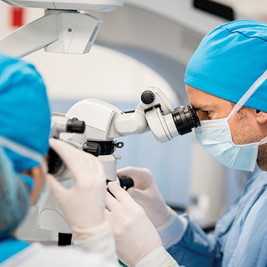
Dr. Mizutani: I have performed surgery on approximately 2,000 cases of cerebral aneurysms. The accuracy and speed of attaching the clip around the neck of a cerebral aneurysm are of utmost importance to determine the surgery’s success or failure. Dissection toward the cerebral aneurysm requires accurate manipulation under microscopy to avoid damage to tissues and bleeding of tiny vessels.
After placing the clip, careful inspection under direct vision is conducted to assure that the clip definitely prevents blood from entering the aneurysm, that the parent artery is not constricted, and that penetrating branches are preserved. Further checking is performed by the ICG* fluorescence imaging.
* ICG: Indocyanine green.
What do you think about the possible utilisation of A.I.M.E. technology for cerebral aneurysm clipping under a microscope?
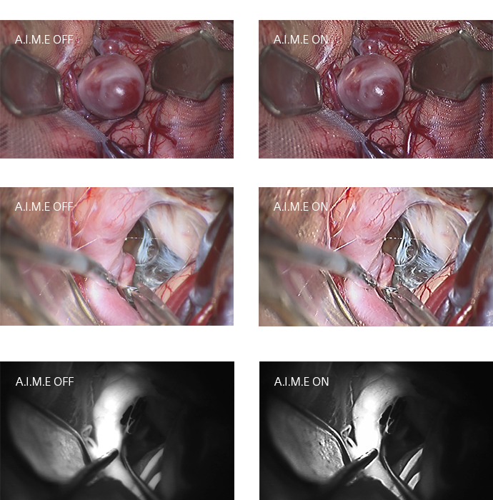
Dr. Mizutani: Compared with the images of clipping preparation I have described before (Photos 1 and 3), those created with A.I.M.E. imaging technology (Photos 2 and 4) seem more clear, and the ‘red colour tone’ is accentuated by the enhancement function of contrast and colour tone. The surgeon actually does not conduct the surgery through a monitor but through the lens of the microscope. Other physicians observing the surgery, however, can see finer tissues and the way in which the surgeon handles the surgical scissors, because the monitor of this system provides very clear images that help them to learn more about the surgery. The system may also be very useful to conduct repeated postoperative checks. Better-defined imaging may even allow surgery using the monitor in future.
When checking with the ICG fluorescence imaging (Photo 5), I noticed that similar clearness was achieved with the contrast enhancement of A.I.M.E. (Photo 6).
Based on these findings, I consider that there are various possibilities of applying A.I.M.E. to procedures performed under a microscope.

How about the expected application of A.I.M.E. technology to other cases?
Dr. Mizutani: When an aneurysm can be approached through the ventricle, a less invasive procedure, a brain endoscopic biopsy can be used. This surgery requires full attention to prevent damage to brain tissues. However, it may be difficult for a surgeon to sense the distance to the target tissue at the time of sample collection for biopsy. For example, in some cases the cerebrospinal fluid is slightly cloudy.
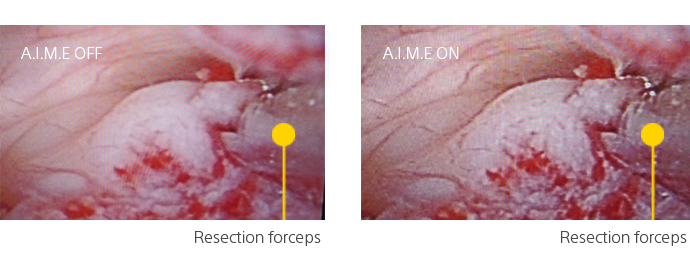
Since the image processed with A.I.M.E. (Photo 8) appears to be better focused than that without A.I.M.E (Photo 7), one can easily grasp the shape of the tissue. A.I.M.E. technology seems to provide clearer vision, even when the surrounding spinal fluid is cloudy, decreasing the risk of surgery.

Profile
Dr. Toru Mizutani
Graduated from the Faculty of Medicine, at the University of Tokyo in 1984. For over 20 years, he has mainly been engaged in the surgical treatment of cerebrovascular disorders, including cerebral aneurysms, carotid artery stenosis, cerebral vascular bypass and benign brain tumors. With his abundant surgical experience, he is considered an expert in this field. Many patients are referred to him from long distances away. He has managed more than 300 cases of dissociative cerebral aneurysm, a world record in the field.


